Outpatient examination
Specialised radiotherapy is available from Monday to Friday in 2 outpatient departments.
Appointments can be made or changed by telephone or email.
What to bring with you?
- Your identity card, social security card
- Your referral documents
- Previous medical documentation relating to your illness
- List of medications you are currently taking
- Results of radiological examinations and their images on DVD
Planned examination
Depending on the type of tumour being treated, planned CT scans are carried out on the day of the outpatient examination or at a later date, if possible in conjunction with the diagnostic examination.
Radiotherapy
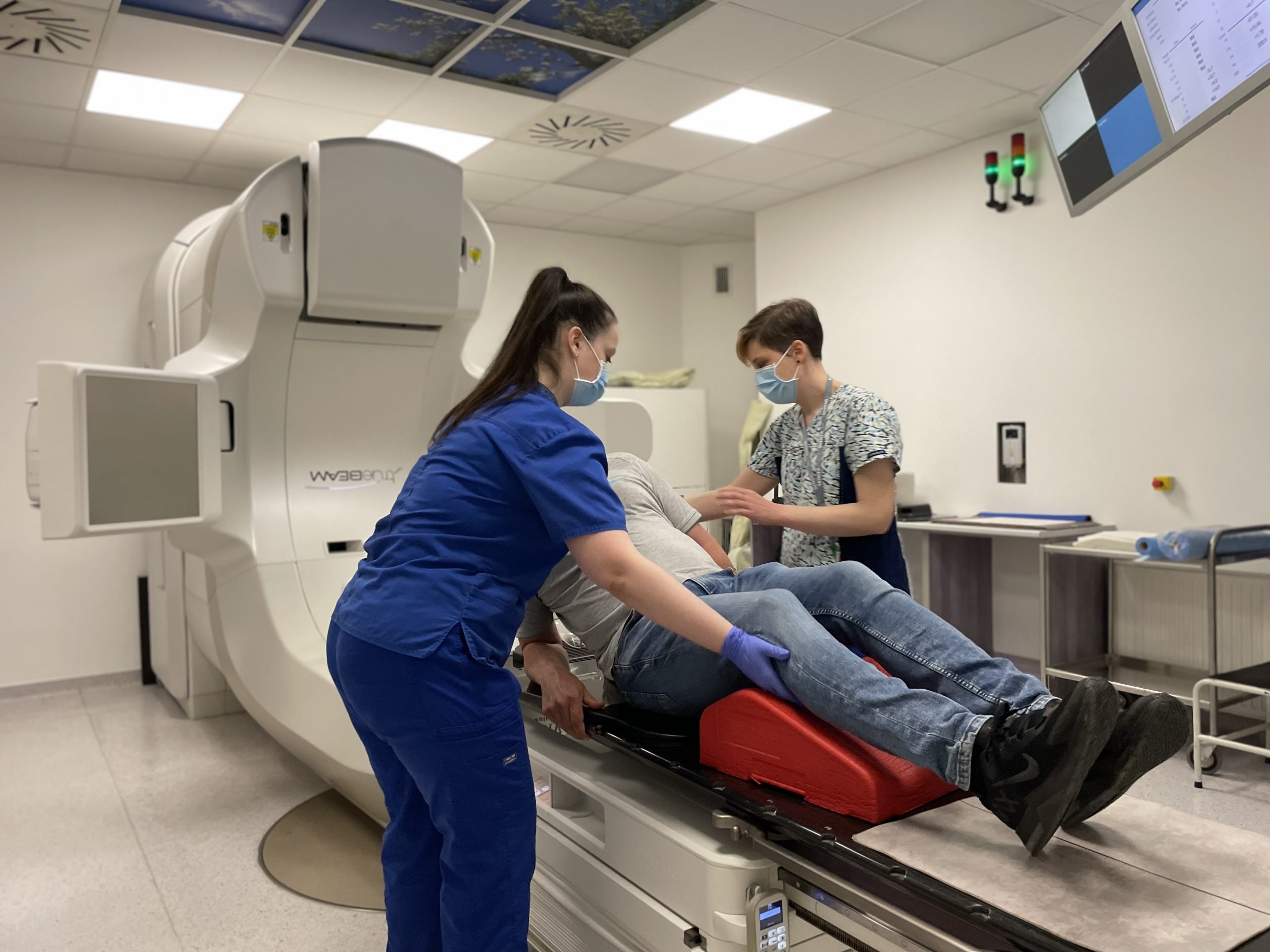
Radiotherapy is scheduled during your outpatient appointment. Treatments are carried out on two modern linear accelerators, with a separate appointment. All our patients are treated on the machine for which they have an appointment. Waiting times may vary between the two machines!
Brachytherapy
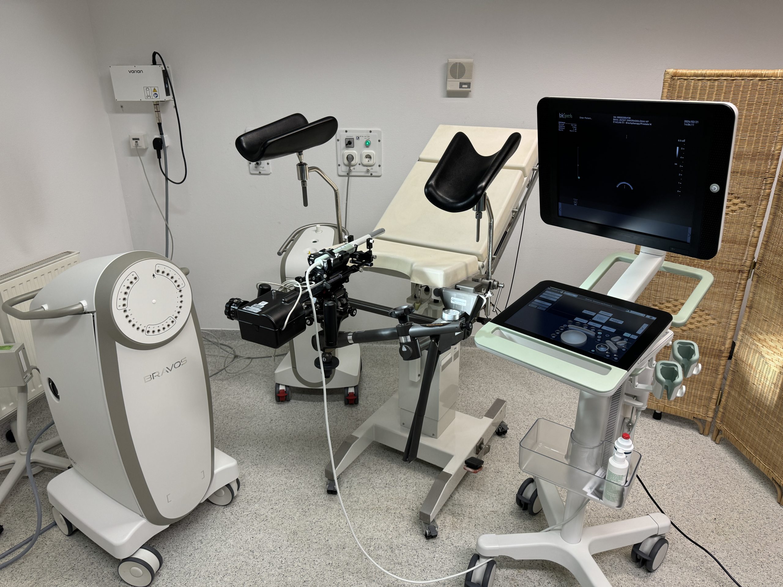
We also provide near-therapy (HDR-AL) for outpatients and inpatients, if the doctor considers it necessary.
Radiotherapy
| Monday – Friday | 8:00 – 16:00 |
PLANNED EXAMINATION
| Monday – Friday | 8:00 – 16:00 |
Brachytherap
| Monday – Friday | 8:00 – 16:00 |
Contact
Treatment Options

Radiotherapy
Radiation therapy is the therapeutic use of high-energy ionising radiation, which inhibits cell division by damaging hereditary material (DNA). However, this process is not selective and affects both intact and cancer cells. However, given that the number of cells preparing to divide or in the process of dividing is usually higher in tumours, tumours are usually more sensitive to radiation than normal tissue. The success of the treatment depends on how the malignant cells react to the radiation, how sensitive they are to it, and how the cells of the healthy tissue surrounding the tumour tolerate the radiation. With careful preparation and the use of modern irradiation techniques, intact organs and tissues can be extremely well protected. At our institution, we treat the following major tumour groups:
- Tumours of the central nervous system
- Stereotactic treatments
- Head and neck tumours
- Lung tumours, thoracic tumours
- Stereotactic treatments
- Mammary tumours
- Abdominal tumours (pancreas, kidney, abdominal lymph nodes)
- Stereotactic treatments
- Urology tumours
- Prostate tumours
- Stereotactic treatments
- Gynaecological tumours (cancer of the uterus, cervix, vulva)
- Colorectal cancers
- Tumours of the skin and soft tissue
- Haematological tumours
- Other malignancies (e.g. bone metastases), in radiotherapy indications
- Stereotactic treatments
- Joint pain (e.g. heel, shoulder, knee)
Brachytherapy
Proximity therapy for gynaecological tumours
Intracavital vaginal high-dose-rate (HDR) proximity therapy
Brachytherapy is a special type of radiation therapy. Vaginal brachytherapy involves the use of radioactive isotopes, which are delivered to the area to be treated using plastic cylinders or cylinders inserted into the vagina. The procedure is well tolerated and requires no special preparation or anaesthesia. The treatment is usually done every other day and does not require hospitalisation.
Proximity therapy for cervical cancer
Proximity therapy is a special radiation treatment. It is carried out using a tiny radioactive substance that is delivered directly into or near the tumour using special hollow proximity therapy devices (applicators) and needles, using remote control. This technique allows a very high radiation therapy dose to be delivered directly to the tumour, while reducing the radiation exposure to the surrounding normal tissue. Radiotherapy for cervical tumours requires a few days in hospital. Treatment is given in two interventions with a one-week break, with a minimum interval of 12 hours between the 2 fractions. Radiotherapy for cervical tumours requires a few days in hospital. During the interventions, ward support is provided by the Inpatient Unit of our Institute.
High dose rate (HDR) prostate brachytherapy given as a boost treatment
A prostate tumour in the high/very high risk group means in part that there is a larger tumour lesion in the prostate, and in part that the tumour has spread beyond the prostate capsule or into the seminal vesicles or is very likely to do so. Today, the vast majority of these patients are treated with a combination of radiotherapy and hormone therapy. Clinical trials over the last decade have shown that in high-risk groups of patients, a more durable local tumour-free outcome can be achieved by increasing the dose to the prostate. In everyday practice, this dose increase can be achieved by modern external beam radiotherapy techniques or by brachytherapy, which involves the delivery of radioactive isotopes to the prostate through needles inserted through a barrier. The radiation physics of brachytherapy allows a very high dose to be generated inside the prostate (i.e. a higher dose to the tumour), while at the same time the dose falls very steeply outside the prostate, sparing the surrounding normal tissues (bladder, rectum), which is not possible to the same extent with external radiation therapy (i.e. reducing the frequency and severity of side effects). The near-therapy is carried out under spinal anaesthesia under sterile conditions in the presence of an anaesthesiologist and a radiotherapist. During the procedure, patients are admitted to the Department of our Institute. The minimum hospital stay is 2-3 days.

Equipment

Irradiation planning
The planned CT scan is performed with SIEMENS SOMATOM Definition AS. Depending on the localisation treated, this is complemented by MR or PET/CT scans. For modern CT-based irradiation planning, the VARIAN ECLIPSE 18.0 planning system is available at our institute. The accurate and faster plotting of the target volume and risk organs is supported by an automatic contouring program based on MIRADA and MVISION artificial intelligence. The connection between the planning system and the linear accelerators is provided by VARIAN ARIA 18.0, a recording and control information system. Comprehensive quality assurance of patient care is supported by VARIAN Mobius. All cancer patients treated at the Institute receive treatment based on individual 3D planning (3D conformal, IMRT= intensity modulated radiation or VMAT= intensity modulated arc radiation).
Radiotherapy
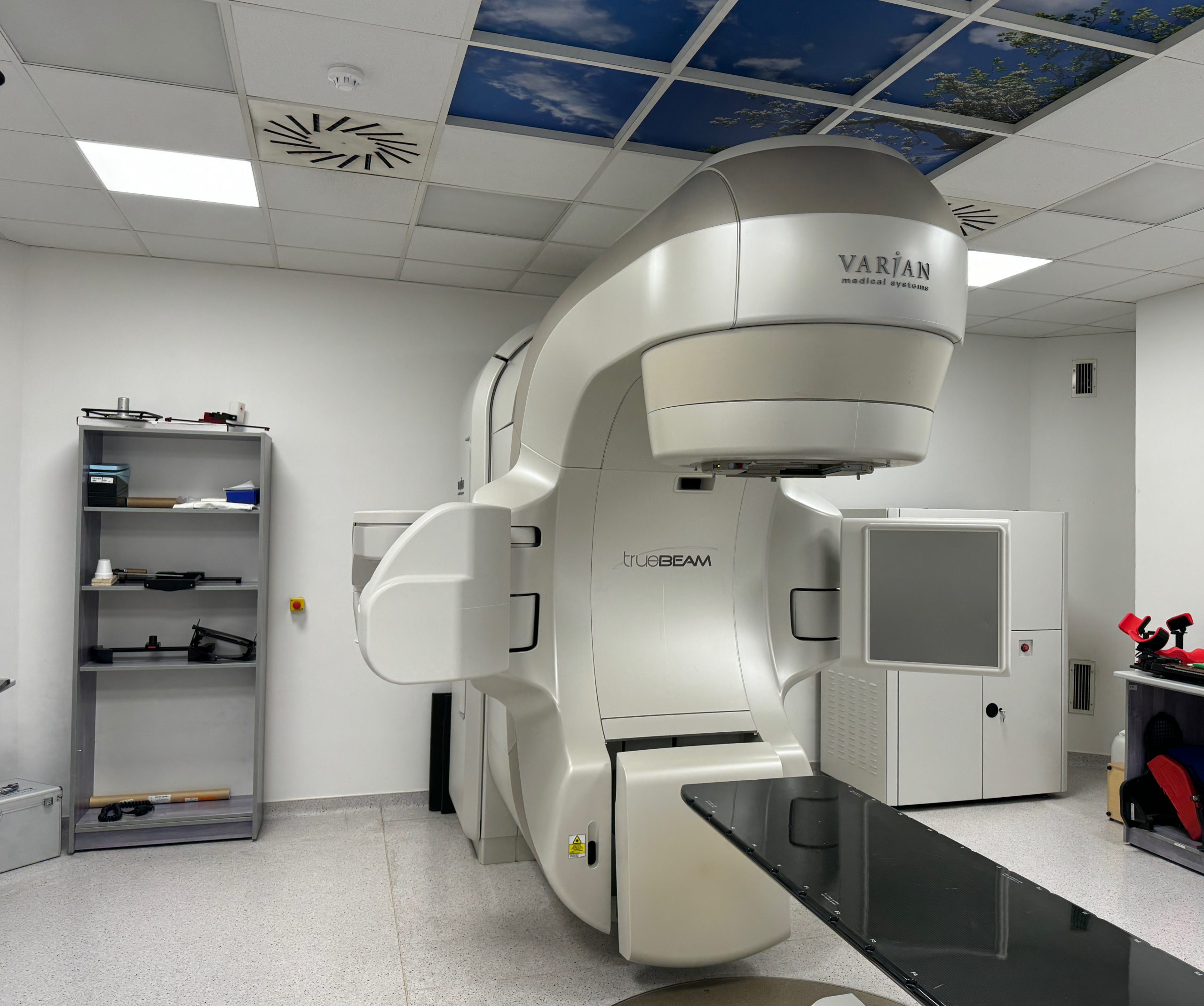
Two modern linear accelerators The VARIAN Halcyon 3.0 is a fast rotating coplanar O-ring linear accelerator. It is treated with 6 megavolt (MV) FFF photon energy. It is equipped with a 4 rpm Dual layer leaf (112 leaf) multi leaf collimator (MLC). The machine is designed to simplify and improve virtually every aspect of image-guided volumetric IMRT and RapidArc therapy technology. It produces high quality kV images with fast data acquisition and advanced reconstruction algorithms. Enhanced IGRT and CBCT image acquisition expands clinical versatility. The VARIAN TrueBeam 3.0 is the latest generation of linear accelerators. In addition to 6, 6 FFF, 10 and 15 MV photon energies, it can also handle 6, 9, 12, 15 and 18 MeV electron beams. It is capable of performing the most advanced arc-based treatments (RapidArc, stereotaxy) with precision and speed. It is equipped with an on-board kilovoltage (kV) imaging system (OBI), which can be used to perform CBCT directly before or during the patient treatment. It can be used to perform image-guided treatments (IGRT) routinely. It can also be used for special imaging: respiration-synchronized MV/kV X-rays, 4D CBCT, Iterative CBCT. And thanks to the extremely high image quality, it can also be used for adaptive radiotherapy following changes in tumour size or shape. Thanks to the built-in respiration monitoring system, it can also deliver breath-hold treatments. With the Advanced IGRT & Motion package, we can also track tumour movement during treatment and correct the setting if necessary during breath-hold and stereotaxic (SABR) treatment.
Brachytherapy
At our institute, we use a high dose rate VARIAN Bravos afterloader with integrated BrachyVision and Vitesse planning system, which allows real-time planning of HDR treatments without CT imaging.
About Us
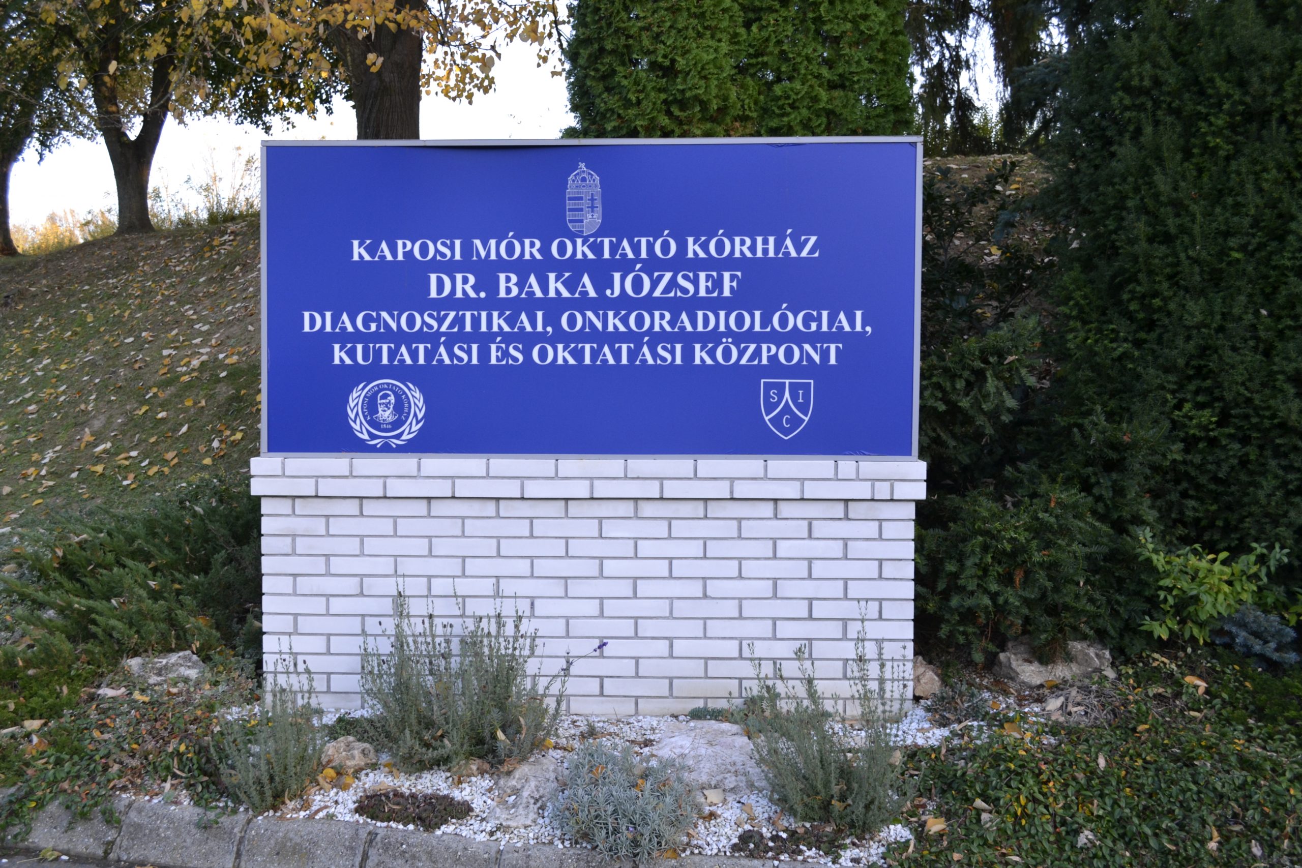
Our institution has been providing radiotherapy treatments within the University of Kaposvár since 2001.Since February 1st, 2018, we have been part of the Complex Oncology Centre of the Kaposi Mór Teaching Hospital of Somogy County. Thanks to the replacement of our machinery in 2015, we can provide our patients with high quality and technological standards of patient care. Since 2017, an automatic contouring program based on artificial intelligence (MIRADA) helps to determine the area to be treated with an accuracy of up to millimetres and to protect organs close to the area to be irradiated during radiotherapy treatments, which further increases the quality of treatment. Complex treatment plans help our patients to enjoy a fuller, more problem-free quality of life by minimising side effects. We can offer our patients a range of modern radiotherapy options for treatment depending on their diagnosis:
- Image-guided radiotherapy (IGRT)
- IIntensity modulated radiotherapy (IMRT)
- Stereotactic radiotherapy (SABR)
- Radiotherapy for joint problems
Coordination
The Diagnostic and Radiotherapy Departments work in close collaboration with our centre. With the PET CT/MR scans performed by Medicopus Ltd., which operates in the building complex, we are able to provide our patients with the necessary imaging examinations (e.g. CT, MR, PET/CT) in a complex and coordinated manner. We can also provide the laboratory tests required for the treatments if necessary. In addition to in-patient and out-patient care, we also offer in-patient ward accommodation in the In-patient Unit, built in 2016.
Oncology patient journey support system
On 1 November 2015, the Kaposi Mór Teaching Hospital of Somogy County established a unified oncology patient journey support system for cancer patients. The aim of the system is to ensure that cancer patients can get diagnosis and treatment as quickly as possible, that they do not circulate in the healthcare system without support, and that their disease can be tracked by their treating physician.


